The Moini et al.'s insect vision chip is a biologically inspired motion sensor [Moini et al. 93, Moini 94, Yakovleff et al. 93]. It is an implementation of the template model [Horridge and Sobey 91] for insect vision.
A simplified model of insect visual system is illustrated in
Figure 3.11. The insects neuro-optical system is
composed of three layers: lamina, medulla, and
lobula or lobula complex. Lamina consists of photodetectors
and automatic gain control circuitry. Medulla contains the small
spatial field motion detectors, in addition to many other complicated
functions. The primary wide-field motion computation is located in
lobula complex. Lobula plate ( a part of the lobula complex) is
also characterized by large directionally sensitive motion detection
(DSMD) neurons. In the template model the motion information is
obtained by thresholding the temporal gradient of the intensity,
![]() , at each pixel. The resulting output indicates
three states: Increase, Decrease, and No-Motion, which can be coded
using two digital bits. This output is then sampled and stored.
Templates are formed by collating the outputs of two contiguous
cells and at two consecutive sampling instants. The templates are then
coded to represent low level motion information. The rest of the
processing which involves tracking of some specific templates is done
using six tracking engines. The location of the tracked templates are
reported off-chip.
, at each pixel. The resulting output indicates
three states: Increase, Decrease, and No-Motion, which can be coded
using two digital bits. This output is then sampled and stored.
Templates are formed by collating the outputs of two contiguous
cells and at two consecutive sampling instants. The templates are then
coded to represent low level motion information. The rest of the
processing which involves tracking of some specific templates is done
using six tracking engines. The location of the tracked templates are
reported off-chip.
The chip has a 1D array of 64 photoreceptors, followed by a
differentiator shown in Figure 3.13. It also
contains RAMs for storing the templates and final results only for
interfacing purposes. Six search-and-track engines have also been
implemented which operate on specified areas of interest in the image.
The chip has been fabricated in a 2 ![]() m CMOS process in an area of
4.5mm
m CMOS process in an area of
4.5mm ![]() 4.6mm. The detectors and analog processing elements only
occupy 1.8mm
4.6mm. The detectors and analog processing elements only
occupy 1.8mm ![]() 0.6mm and the rest is dedicated to digital
processing modules.
0.6mm and the rest is dedicated to digital
processing modules.
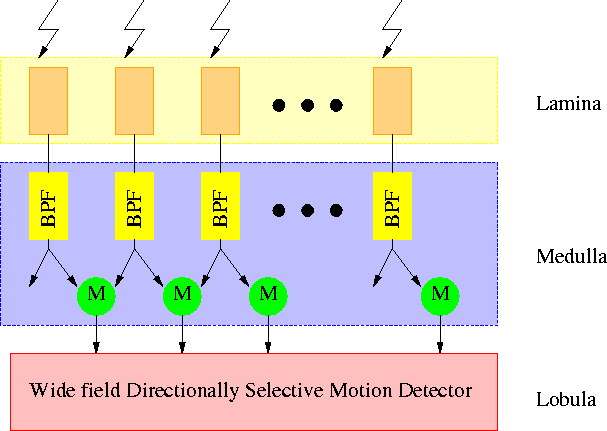
Figure: A simplified block diagram of insect visual system used in
Moini et al.'s insect vision chip.
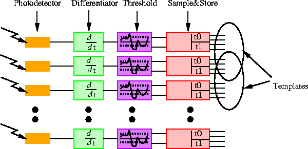
Figure: Formation of templates in Moini et al.'s insect vision chip.
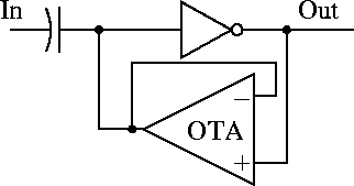
Figure 3.13: Schematic of Moini's differentiator.