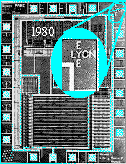
Richard Lyon's optical mouse [Lyon 81a, Lyon 81b, Lyon and Haeberli 82] is one of the earliest smart vision sensors designed and implemented on silicon. Apart from the specific design approach used for the optical mouse, Lyon also pointed out some of the very fundamental aspects of architectural design methodologies for VLSI smart sensors, i.e. using very simple and conservative device models, design rules, analog and digital circuits, and timing techniques. This methodology has been considered in almost all the analog VLSI chips comprising large arrays of analog circuits to date, and is not going to change unless VLSI processes mature to a highly uniform and reliable level.
Lyon's eye is basically a digital motion detection chip. Photodetection is performed using diffusion junction diodes in p-substrate in an NMOS process. Photocurrent is integrated over time on a capacitor, and transduced to a digital signal by a simple inverter. The time that takes for the photocurrent to charge up the input capacitance depends on the light level. Therefore, if the neighboring cells laterally inhibit each other, a cell exposed to a brighter pattern will have a "high" state earlier than other cells. Thus, it can inhibit neighboring cells from firing to a high state. By tracking the location of winner cells at consecutive times, the movement of the input image, which is the fixed pattern of the mouse pad, can be determined. The references discuss the characteristics of the lateral inhibition when used with different neighborhood coverage radii. The algorithm used for detecting motion is intuitive and simple, and is based on tracking the particular pattern of the mouse pad.
The chip was designed in a 5 ![]() m NMOS process. A 4
m NMOS process. A 4 ![]() 4 array of
photodiodes, digital circuits for lateral inhibition, and other
digital circuits for interfacing were implemented in an area of
3.5mm
4 array of
photodiodes, digital circuits for lateral inhibition, and other
digital circuits for interfacing were implemented in an area of
3.5mm ![]() 3.5mm.
3.5mm.
Figure 3.1: Microphotograph of Lyon's eye.